Case Study Function in Implantology
Function in Implantology
A case report by Dr. Umut Baysal – Implant treatment based upon real jaw movements
Function and aesthetics in implantology - The succes of a treatment with implant-supported dental restorations is determined not only by the aesthetic rehabilitation, but more so by functional durability
Introduction
Measuring the mandible movements is an essential step in prosthetic rehabilitation after the loss of a tooth. The use of implants is an excellent treatment option for an edentulous jaw, with many advantages in comparison to conventional methods.
This report addresses the importance of instrumental jaw movement registration and its application for adjusting the articulator. The zebris system is used to record the jaw movements digitally. Especially in a case were all support zones are lost, a redetermination of the jaw relation is of great significance. The precision of the registration is primarily responsible for the accuracy of the new occlusion. Due to the lacking flexibility and buffer effect of an osseointegrated implant, the requirements for occlusal precision of implant-supported restorations are exceptionally high. In function and parafunction, occlusal forces impact on the implant without any periodontally distributed, reflexive cushioning effect.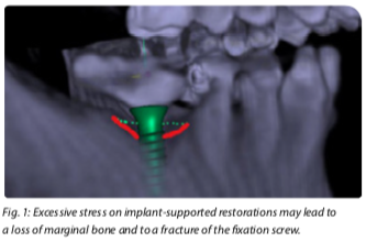
Forces impacting on the implant due to a faulty design of the occlusion may lead to excessive stress and biological complications such as tensional pain, irritation of the soft tissue surrounding the implant, micro-fractures of the crestal bone and even a premature loss of the osseointegration. Thus, it is wise and necessary to use especially precise techniques to determine the maxillo-mandibular relationship during clinical implementation. By including the jaw movement records in the implant planning, the desired outcome of the treatment is obtained with much greater certainty. The objective is to create a functionally harmonious correlation of temporomandibular joint, muscles and occlusal stabilization.
Patient Case (Casuistry)
A 50-year old male patient first presented at the end of the year 2017. The maxilla was edentulous aside from teeth 13, 12, 22 and 23. In the mandible, only the teeth 34 and 43 remained. As a restoration, the patient had previously received simple clasp dentures for both jaws. The clinical degree of tooth loosening was 2-3. After a detailed consultation, we listed the following requirements to a future dental restoration:
- removable (extendable)
- design without a palatal plate
- implant-supported
- economical price
Together, we decided on two removable telescopic dentures. The design for the maxilla restoration was planned without a palatal plate.
Diagnostics and Planning
A CBCT (threedimensional imaging) was ordered. The remaining teeth left for the interim served to support the scan template. Later, they were also meant to support the drill template. Due to their degree of loosening, a wide basal support was provided.
zebris Measurement
At this time, the patient‘s jaw movements were first measured with the zebris JMAnalyser+ jaw registration system (Schütz Dental, Rosbach/Germany) in order to have a better knowledge of the patient‘s jaw movements and possible therapeutic measures, e.g. a bite elevation. zebris is suited to be used in an analogous or in a digital workflow. A measurement takes about 15 minutes and can be integrated into the day-to-day practice routines.
Implant Planning
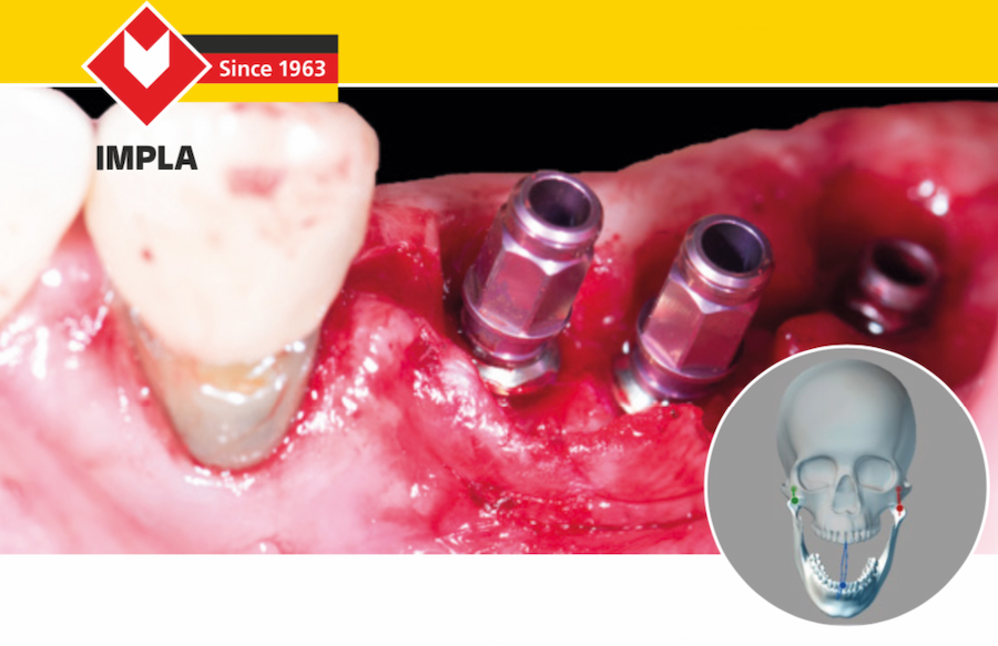
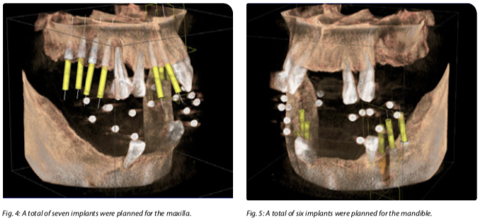
A total of seven implants were planned for the maxilla (fig. 4) and a total of six implants were planned for the mandible (fig. 5). The reduced bone height in the area of the molars necessitated a surgery with simultaneous internal sinus lift. Due to the indication, the use of both IMPLA Cylindrical Cone Connection implants and IMPLA Micro Retention implants (all implants by Schütz Dental GmbH, Rosbach/Germany) was planned for the maxilla as well as for the mandible. The open system Sicat Implant by company Sicat was used for preplanning.
Implantation
Maxilla
Implantation was carried out under local anesthesia and in two sessions. After a crestal incision and one-sided mesial relief, the pilot drillings were placed with help of a drilling template.
Preparation of the implant site
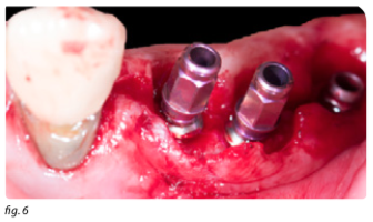
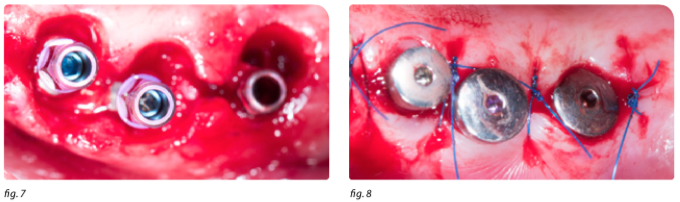
Osteotoms were used to prepare the implant sites in the maxilla, tooth positions 16, 15, 14, 21, 24, 25 and 26. The implantation in positions 16, 25 and 26 was carried out with a simultaneous sinus lift (fig. 6, 7 and 8).
Mandible
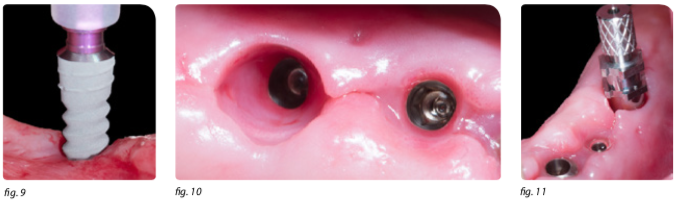
The mandible was prepared in accordance with the prescribed drill protocol with help of a pilot drilling template. The implants were inserted without the use of a drilling template (fig. 9). After 8 weeks, an impression of the implants was taken with impression posts and in the open tray impression technique. This impression was used for the bite registration and for the production of a model (fig. 11). Fig. 10 shows the irritation-free gingiva around the implants.
Bite Registration
For a complex prosthetic rehabilitation, it is advisable to program the articulators to the patient‘s individual and specific needs. When using average values for programming the articulators, deviations of a magnitude of about 300 μm can not be avoided.
zebris Measurement
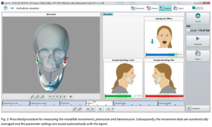
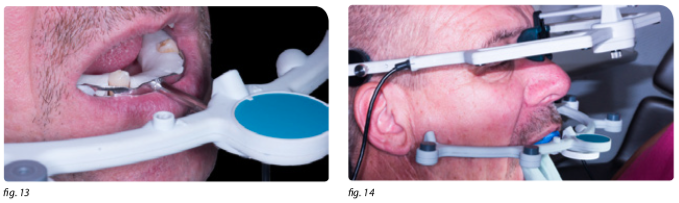
zebris JMAnalyser+ System (Schütz Dental GmbH, Rosbach/Germany) was used for the measurement in this case. The zebris system is an ultrasound-based axiography system. It lets the user determine all important parameters necessary for programming the articulator in a quick, precise and efficient way. zebris measures the propagation time of ultrasound impulses which are transmitted by modules integrated in the lower jaw sensor and which are received by microphones in the face bow.
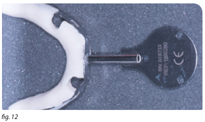
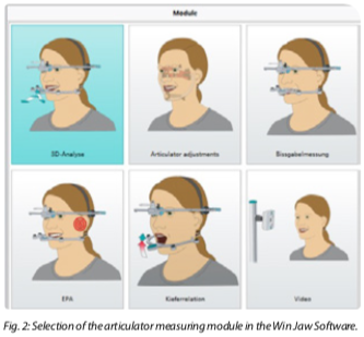
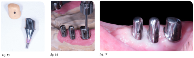
For set up, the face bow is attached and the lower jaw sensor is fixed in the mandible with help of the prepared occlusal tray (fig. 12, 13). The registration of the maxilla is also carried out with the lower jaw sensor. It is fixed to the maxilla with a carrier (fig. 14). The new software facilitates the analysis and documentation of an arbitrary number of movements. The system records all six degrees of the lower jaw mobility. The user can choose between the main modules „articulator“, „function“ and „jaw relation“. The software is operated intuitively and the user is guided through the recording procedure with short instructions and animations. In our case, we used the modules „function“ and „articulator“. After the measurements are finished, the results are exported in PDF-format for individual programming. A report is available for the three most common articulator systems (KaVo, Artex and SAM). With this data, the dental technician can program the digital articulator individually.
Prosthetics
The determination of the vertical and horizontal jaw relation is particularly signifcant in cases where a removable denture is produced for a patient with an edentulous jaw or with little remaining dentition and a concomitant loss of natural support zones. Aside from an overload of the jaw and the jaw joints, an incorrect vertical dimension can lead to problems with lip closure, pronunciation and aesthetic appearance. The registration after implantation is an excellent possibility to circumvent those problems which may result from an edentulous jaw. Therefore, the osseointegrated implants are used as a support when determining the vertical jaw relation. Regardless of the registration technique, an implant-supported deter- mination of the jaw relation facilitates an excellent fixation, even in functional motion.
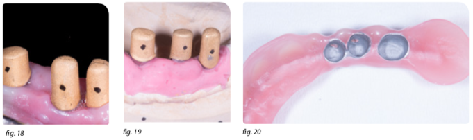
After the dental laboratory has been provided with all pertinent data, the dental technician can start designing the denture. In the following case, we manufactured a telescopic denture without palatal plate. The primary parts were designed with the Tizian Creativ RT CAD Software and milled with the Tizian Cut 5.2 milling machine, both by Schütz Dental GmbH (Rosbach/Germany), see fig. 15. After fitting the primary parts, we prepared an impression for a second model (fig. 16 and 17).
We decided against a ceramic veneered bridge, as we believe that, with a loss of soft tissue, such a bridge with pink ceramic is not superior to the acrylic version. The galvanic copings were bonded intraorally for a „passive fit“ (fig. 18) which facilitated a friction-free integration of the denture. The secondary crown shown in the picture was cast conventionally, as an ultra-precise fit was not required (fig. 19 and 20).
Result
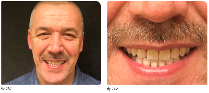
The final photograph shows a happy patient who significantly gained in quality of life after wearing a clasp prosthesis for many years (fig. 21.1).
Summary
An exact determination of the jaw relation that includes the recording and transmission of the real jaw movements is a major step towards rehabilitation after the loss of teeth. With the zebris system, the mandibular hinge axis can be determined with sufficient accuracy. This system features a good reproducibility of the mandibular hinge axis‘ location. The necessary data to program the articulator completely are recorded and transmitted to the dental lab without effort.
Author

Dr. med. dent. Umut Baysal
Dentist for Implantology
Praxis am Nordwall
Nordwall 2, 57439 Attendorn
Mail: u.baysal@icloud.com
Download this case study as PDF file:
Please note: This case study is not an instruction leaflet. Please adhere to the instructions provided with the material and/or equipment. The responsibility for the treatment remains with the attending dentist.


Tactile scans ... are worth the effort!

Bleaching with "Bleach’n Smile“

Case study: Cervical Fillings with PrimeBond7 and Capo Slow Flow
